Skip to content

Courses/Postdegree in Ultrasound Imaging Course

Postdegree in Ultrasound Imaging Course

2,600.00 USD

100% finance available with
  • Level

    Advanced

  • Language

    Spanish/English

  • Estimated duration

    6 months

  • Recommended input

    10 class hours a week, flexible itinerary to suit your pace

  • Subscription

    Valid for 12 months

  • Accreditation

    EUNEIZ university · 16 ECTS

  • Accreditation fees not included

Clinical context

Ultrasound allows you to assess anatomical structures in real time and provide information for clinical examination. Its use requires anatomical knowledge, the obtaining of quality images, and the relating of the findings to the patient's situation.
Transversal training in ultrasound allows you to:
  • ✓Perform systematic examinations in different anatomical regions.
  • ✓Recognize normal images, anatomical variants, and relevant pathological findings.
  • ✓Improve spatial orientation and the identification of structures.
  • ✓Incorporate ultrasound imaging into clinical reasoning within the scope of skills of each professional.

What is this course?

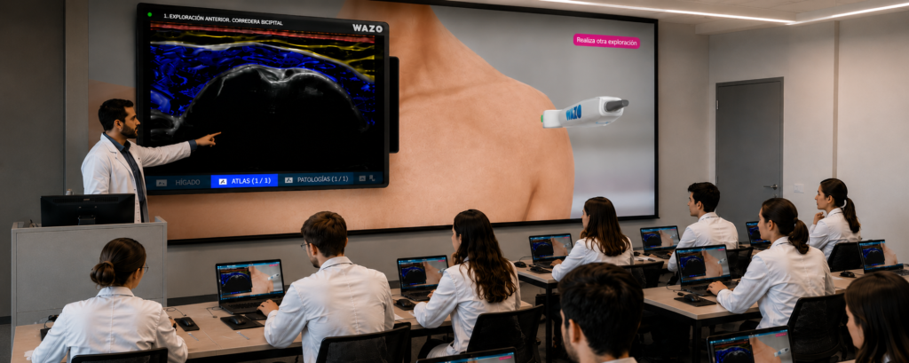
Wazo® University Ultrasound Postdegree Certificate, with 400 course hours and 16 ECTS, developed in collaboration with the EUNEIZ University. 100% online course available through the Wazo® Campus that combines theoretical training, guided practices, interactive simulators and theoretical and practical assessment.

Training objective

To develop the necessary anatomical, technical, and clinical knowledge to carry out systematic ultrasound examinations, recognize relevant structures and findings, and incorporate the image information into clinical reasoning, through progressive learning and simulation.

Training areas

Abdomen Neck and thyroid Breasts Soft tissues and abdominal wall Male genitalia Doppler Musculoskeletal system Lumbopelvic region and pelvic floor

Clinical approach

Abdomen: examination of blood vessels, liver, bile ducts, pancreas, spleen, kidneys, and bladder. Neck and thyroid: recognition of the suprahyoid and infrahyoid regions, the thyroid gland, and the cervical lymph nodes. Breasts: normal anatomy, examination technique, pathologies, and correlation with BI-RADS. Soft tissues: assessment of the skin, subcutaneous tissue, and abdominal wall. Male genitalia: ultrasound examination of the scrotum and penis. Doppler: hemodynamic fundamentals, waveform analysis, and technical adjustment. Musculoskeletal: anatomical recognition and examination of the shoulders, elbows, hands, hips, knees, and ankles. Lumbopelvic: ultrasound assessment of the abdominal and lumbar muscles, pelvic floor, and diaphragm.

What does it include?

  • ✓Nine modules that cover the basic principles and regional applications of ultrasound.
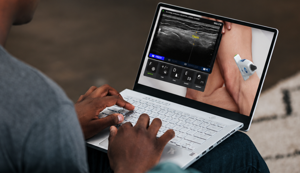
  • ✓Pre-recorded theoretical classes and examination videos with real models and ultrasounds.
  • ✓Demonstrations featuring a triple synchronized display of the environment, the examination area, and the ultrasound screen.
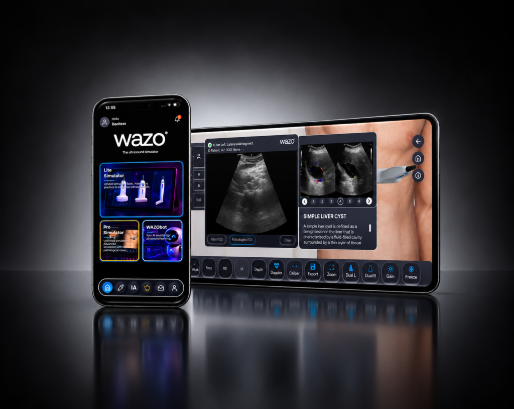
  • ✓Online clinical simulators available 24 hours a day during the training period.
  • ✓Ultrasound atlases, images, downloadable materials, and examination protocols.
  • ✓Virtual clinical case studies to relate anatomy, image, and clinical context.
  • ✓Theoretical exams and practical tests on the identification of structures in a gamified environment.
  • ✓Academic assistance through Wazo®'s specialized ultrasound AI.
  • ✓Progress monitoring, activity log, and assessment results.
  • ✓Access to the content for a year and academic and technical support.

What will you learn?

  • ✓To understand the physical principles of ultrasound and optimize the equipment settings.
  • ✓To apply examination protocols for abdominal, cervical, thyroid, breast, and soft tissue examination.
  • ✓To recognize the structures of the male genitalia and their main ultrasound findings.
  • ✓To understand the basic principles of the Doppler and vascular flow analysis.
  • ✓To examine the musculoskeletal regions of the upper and lower extremities.
  • ✓To assess the abdominal, lumbar, pelvic floor, and diaphragm muscles through ultrasound within the RUSI approach.
  • ✓To relate ultrasound findings to clinical assessment and recognize examination limitations.

How will you learn?

  1. 1Learn: physical principles, anatomy, and ultrasound technique through structured content.
  2. 2Practice: follow step by step examination demonstrations and protocols.
  3. 3Compare: use atlases and images to recognize ultrasound structures and patterns.
  4. 4Examine: apply the content to virtual clinical case studies and scenarios.
  5. 5Repeat: train on simulators during the access period to reinforce skills.
  6. 6Assess: complete questionnaires and gamified practical tests with a results log.

Postdegree in Ultrasound Imaging Course
Course

Request a demo and we'll design a tailored plan for your service or university.