Skip to content

Simulators/Echocardiography

Echocardiography

59.00 USD

100% finance available with
  • Level

    Advanced

  • Language

    Spanish/English

  • Subscription

    Valid for 12 months

Clinical context

The assessment of a patient with dyspnea, hypotension, or hemodynamic instability requires the incorporation of the clinical examination with information on the structure and function of the heart. Clinical echocardiography provides bedside imaging to guide this assessment.
  • ✓To recognize the cardiac chambers and assess their relative size.
  • ✓To visually estimate the global systolic function of the left ventricle.
  • ✓To identify pericardial effusion and recognize signs of possible hemodynamic repercussion.
  • ✓To recognize increased size of the right ventricle.
  • ✓To explore the inferior vena cava and interpret the findings in the clinical context.
  • ✓To incorporate ultrasound findings into the initial assessment of shock.

Training areas

Parasternal window Apical window Subcostal or subxiphoid window Cardiac chambers Septa and valves Pericardium Inferior vena cava

Clinical approach

Anatomy and cardiac views: orientation of the image and recognition of the structures in the different projections. Inferior vena cava: assessment of its diameter and respiratory variation as supplementary information for hemodynamic assessment. Pericardium: recognition of pericardial effusion and signs of possible tamponade. Left ventricle: recognition of normal, reduced, and gravely reduced global hyperdynamic function. Right ventricle: identification of increased size and revision of signs of overload, including the D sign and the McConnell sign. Shock: incorporation of findings

What does it include?

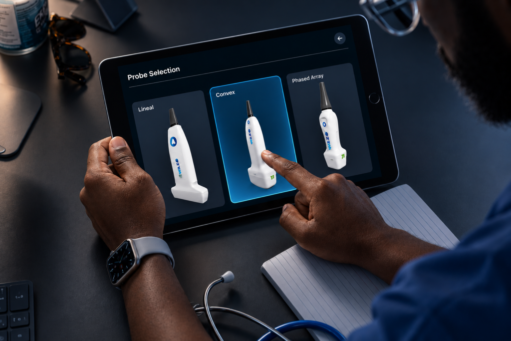
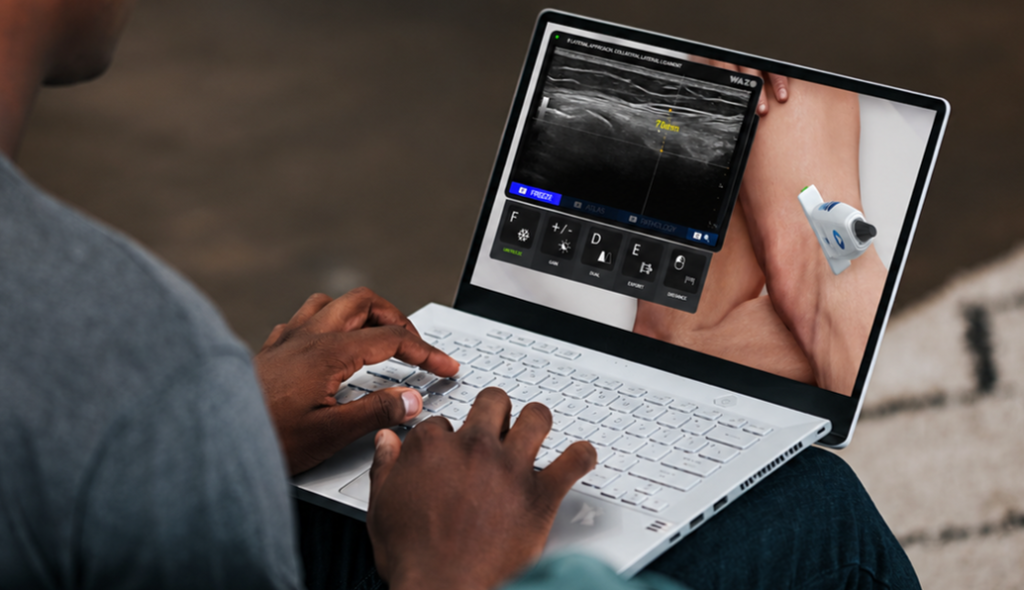
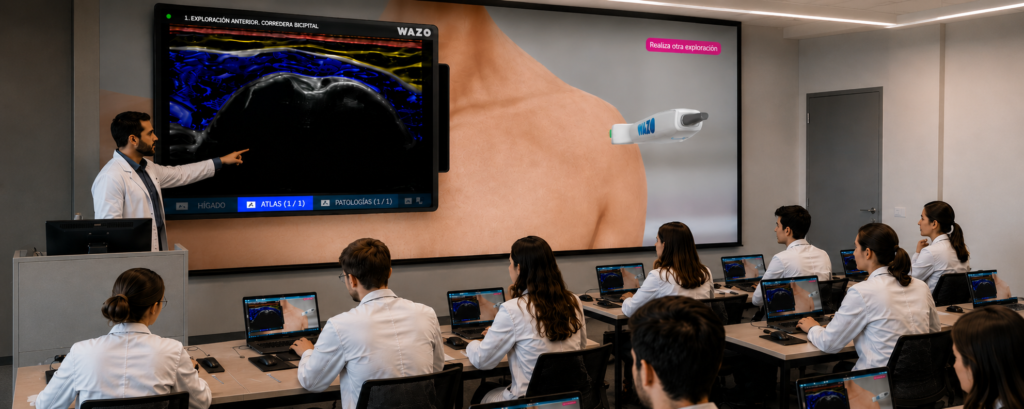
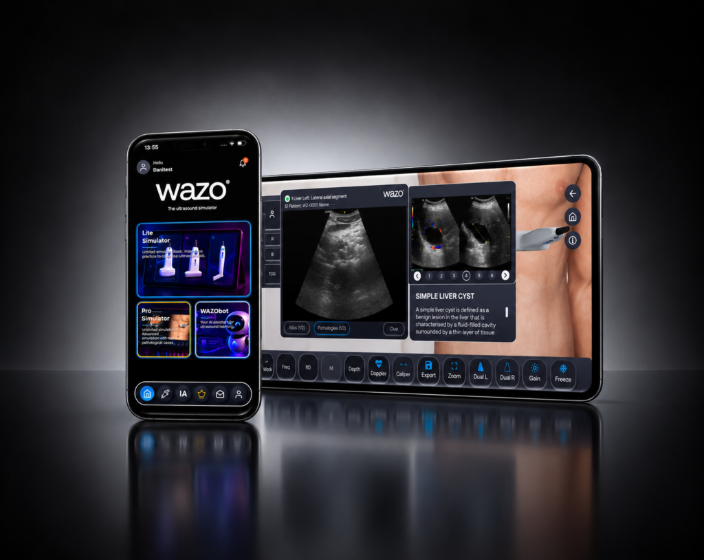
  • ✓Practical training in the Wazo® simulator.
  • ✓7 ultrasound approaches to learn how to perform examinations.
  • ✓14 ultrasound atlases and 100 anatomical structures to reinforce the identification of anatomy.
  • ✓18 clinical case studies to relate the findings to clinical situations.
  • ✓Progressive learning through the relationship between anatomy, image, and clinical context.

What will you learn?

  • ✓To recognize the parasternal, apical, and subcostal windows and the major cardiac projections.
  • ✓To identify chambers, septos, valves, and anatomical references in the images.
  • ✓To assess the size and respiratory variations of the inferior vena cava.
  • ✓To recognize pericardial effusion and ultrasound signs of tamponade.
  • ✓To visually clarify the global systolic function of the left ventricle.
  • ✓To identify dilation of the right ventricle and signs of overload.
  • ✓To relate the findings to shock patterns and the RUSH protocol approach.

How do you learn?

  1. 1Learn: the fundamentals, indications, and anatomy of clinical ultrasound.
  2. 2Practice: orientation and examination of the approaches through simulations.
  3. 3Compare: anatomical images and ultrasound with the support of the atlases.
  4. 4Integrate: the relationship between ultrasound imaging and clinical situation.
  5. 5Explore: clinical case studies
  6. 6Reinforce: revision of the structures and image patterns.

Echocardiography
Simulator

Request a demo and we'll design a tailored plan for your service or university.